
Uterine fibroids (fibroids) are known by many names. These are often called leiomyomas, myomas, uterine myomas, and fibromas. Whatever the name, fibroids are rubbery tissue masses that develop in the muscular portion of a woman’s womb.
Fibroids have different classifications. These growths come in different sizes, shapes, and colors. They can be grow inside or outside the uterus of a woman.
Fibroids are usually identified through radiographs, transabdominal and transvaginal scans, CT scans, and MRI.
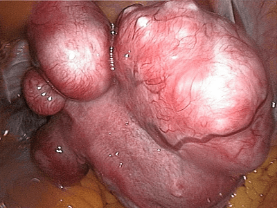
How uterine fibroids look like?
CLICK HERE TO GET RID OF YOUR FIBROIDS NATURALLY WITHOUT SURGERY
Fibroids are classified according to their location (inside or outside the uterus).
Submucosal –
This is the least common type which accounts for only 5% of all fibroids. These type of fibroids grow in the endometrium. The endometrium is the thin innermost layer or lining inside the uterus. They grow toward the peritoneal cavity, are broad-based (sessile) or attached to the surface by a stalk (pedunculated). Submucosal fibroid can cause profuse bleeding and are often linked to fertility problems.
Subserosal–
This type is located underneath the serosal surface, which is a thin layer that lines the cavities of the body and serves as covering of the outside surface of organs. They grow outward the peritoneal cavities, that space within the belly that contains the liver, intestines, and the stomach. They can extend to the broad ligament. Subserosal fibroids may not cause bleeding but it can lead to intense pressure. Sufferer could be subjected to a of pain.
Intramural –
This is the most common of type of fibroids. These growths are found in the uterus’ myometrium, which is the thickest mid-layer of the uterus. It consists of a smooth muscle that contracts during menstrual period. It can lead to the removal of the endometrial lining.
It can change the shape of the uterus. There are no manifestation of the symptoms early on. However, these fibroids can cause infertility because of the compressed fallopian tubes.
Doctors use the classification above to identify the fibroids. However, the presentation depends on the number of lesions, size, and location of the fibroids.
Fibroids are thick masses of normal muscle cells. The surfaces range from white to tan colors and show a whorled trabecular pattern. Trabecular pattern consists of stress lines along the bone. The appearance of fibroids often changes because of degenerative alterations.
Seen through a microscope, fibroids exhibit whorled, anastomosing fascicle which appears uniform with the shape of a spindle. The center of the fibroid is elongated with finely spread chromatin.
Uterine fibroids are benign tumors. They are not usually detected immediately as the condition show no visible symptoms. These tumors are usually accidentally identified because of imaging and/or testing for other illnesses. Fibroids are often easy to recognize, but when their degeneration starts they would appear differently and come in many different and weird shapes and sizes.
Knowing the appearance of fibroids is significant in the correct identification the condition. Doctors who suspect the presence of these fibroids in the body of the patient would recommend tests to accurately identify or rule ou the condition.
